Image from Dreamstime
Foot Type and Low Back Pain
By: Dr. Emily Splichal
This information is from Dr. Splichal’s eLearning continuing education course:
Biomechanics of Low Back Pain. See the full course to learn more. The course includes great graphics and video for assessment.
Low Back Pain from the Ground Up
It is important to understand basic foot anatomy and function in order to learn how deviations from functional norms can occur in the foot and ankle, translating up through the knee, hip, and low back. These deviations can contribute to the different foot types and specifically how each foot posture will affect the biomechanics of low back pain.
In this article you will review the three most common foot types encountered by the health-fitness professional. These different foot types can be responsible for a variety of proximal biomechanical imbalances affecting the knee, hip and lower back. If these biomechanical imbalances are not addressed, they can lead to kinematic compensations and an increased risk of injury.
As you review each foot type in detail, try to visualize the compensations and how you, as a health-fitness professional, can use stretching and strengthening programs to help reverse these postural deviations and biomechanical compensations.
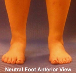
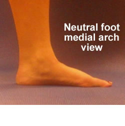
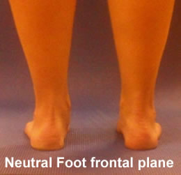
Neutral Foot
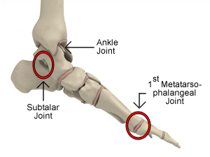
The neutral foot is characterized by optimal range of motion in the ankle, subtalar (STJ) and 1st metatarsal phalangeal joint (MPJ). The neutral foot allows every step you take to move through the sagittal plane without deviation. The neutral foot would not be responsible for biomechanical imbalances proximally, and should therefore present with optimal knee, hip and pelvis function.
The basic characteristics of a Neutral Foot include:
Toes are not abducted on the forefoot
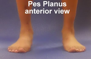
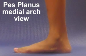
Pes Planus Foot Type
Over-pronation, or a pes planus foot type is perhaps the most common of all foot biomechanical problems encountered by the fitness professional. Although we all pronate during bipedal locomotion, foot pronation that exceeds 25% of midstance is abnormal and will impact proximal kinematics. Many pathological conditions are associated with over-pronation including bunions, medial knee pain and low back pain, which is why correction of this foot type is of particular importance to the health-fitness professional.
The basic characteristics of a Pes Planus foot include:
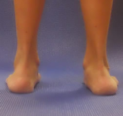
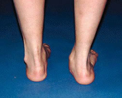
Biomechanics of a Pes Planus Foot
Pes planus foot, increased internal leg rotation and low back pain:
With the foot acting as the interface between the ground and the rest of the body, and since most motions of the foot are in a closed chain environment, motion of the foot must somehow be translated more proximally through the leg to the hip and lower back. This translation in foot motion is in the form of tibial internal and external rotation. The direction and degree of tibial rotation is dictated by the subtalar joint or more specifically the talus.
The interaction between the leg and foot is often described as a mitered hinge or a screw. In a pes planus foot the subtalar joint is in an everted position and is therefore associated with internal leg rotation. This coupled motion can best be remembered by whatever the calcaneus does, the leg does the opposite motion.
Going back to our screw analogy, if we look at the left leg and foot, as we screw the leg internally, the medial arch collapses and the calcaneus everts. As we untwist the screw or externally rotate the leg we could visualize how the foot begins to supinate, the arch raises and the calcaneus inverts.
So how does increased tibial rotation cause low back pain?
Bilateral (both sides) pes planus and Low Back Pain:
In a client presenting with a bilateral pes planus foot posture, we would want to appreciate the prolonged internal rotation of the tibia and femur. As both femurs internally rotate, the supporting ligaments of the hip joint force the pelvis into an anterior tilt position. The anterior tilt position can lead to a myriad of biomechanical problems including altered reciprocal inhibition and increased shear forces to the lumbar vertebrae.
Unilateral (one side) pes planus and Low Back Pain:
A client with unilateral pes planus must be further assessed for possible limb length discrepancy. This was described in detail in the Functional Foot and Ankle course and is reviewed again in the Biomechanics of Low back Pain course. The biomechanical impact of a unilateral pes planus or limb length discrepancy plays on low back pain is through a frontal plane imbalance and stress to the sacroiliac (SI) joint.
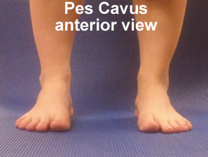
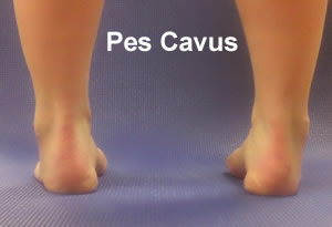
Pes Cavus Foot Type
Although the flat foot, or over-pronated pes planus foot type is considered more significant in terms of foot dysfunction and biomechanical imbalances, the high arched, or pes cavus foot type can surprisingly cause more significant pathology.
The basic characteristics of a Pes Cavus foot include:
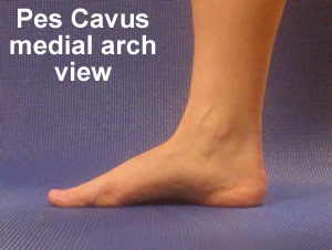
Biomechanics of a Pes Cavus Foot
Pes Cavus foot, uneven weight distribution and low back pain:
Since the pes cavus foot lies in a more supinated or inverted position, this foot is associated with uneven weight distribution to the lateral aspect of the foot. This uneven weight distribution to the outside of the foot will require increased stabilization and activation from the peroneals. Since the peroneal fibers run along the lateral aspect of the leg and into the iliotibial (IT) band, increased tension in the peroneals must place greater tension on the IT band. Increased tension in the IT band will cause tension into the tensor fascia latae muscle and an anterior tilt of the pelvis.
Pes Cavus foot, poor shock absorption and low back pain:
Another important characteristic of the pes cavus foot type is that it is a poor shock absorber. When we walk our foot moves from heel strike to propulsion, with a concomitant supination into pronation, and back to re-supination. This coordinated movement of the foot is intelligently designed to absorb shock and allow us to walk on uneven terrain. In a pes cavus foot, the foot does not adequately pronate during midstance and therefore does not absorb all ground reaction forces. This means ground reaction forces must be taken up by our tendons, ligaments, and bones. It is for this reason that the pes cavus foot type is often associated with Achilles tendonitis, IT band bursitis, and possible SI joint dysfunction.
About the Author:
Dr Emily Splichal, Podiatrist and Human Movement Specialist, (DPM, MS, MPH, NASM-CES, NASM-PES, NSCA-CPT) is the Founder of the Evidence Based Fitness Academy. With over 10 years in the fitness industry, Dr Splichal has dedicated her medical career towards studying postural alignment and human movement as it relates to foot posture and foot strength. Dr Splichal is expert lecturer and TV personality with appearances on Oprah Winfrey, The Today Show & Good Day NY. Dr Splichal is sought after for her expertise in barefoot training, foot health, and postural alignment.
To learn more about the Evidence Based Fitness Academy and Dr Emily Splichal please visit www.evidencebasedfitnessacademy.com
Submitted:
October 1, 2014
By: June Chewning
Fitness Learning Systems
513 367-1251
June@FitnessLearningSystems.com